Acute pulmonary embolism
High-risk pulmonary embolism (One of the following)
Cardiac arrest
Obstructive shock


90
Systolic BP < 90 mmHg
OR
Vasopressor required
to achieve a BP >_90 mmHg despite adequate filling status
And
End organ hypoperfusion
(oliguria, alter mental status or increase serum lactate)
Persistent hypotension
90
40
mmHg
Systolic BP < 90 mmHg
OR
systolic BP drop >_40 mmHg,
lasting longer than 15 min and not caused by new-onset arrhythmia, hypovolaemia, or sepsis
None of above ?: See Not high risk.
Suspected PE with hemodynamic instability


Probability for PE
Well's score
Classic Well's score
Low
Intermediate
High
PE unlikely
PE likely
Simplified Well's score
Low
Intermediate
High
PE unlikely
PE likely
Echocardiography
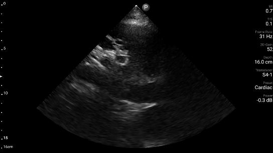
Parasternal long axis (PLAX)

In the parasternal long axis view, there is an enlargement of the right ventricle (the structure of the right chamber of the heart located at the front adjacent to the sternum).
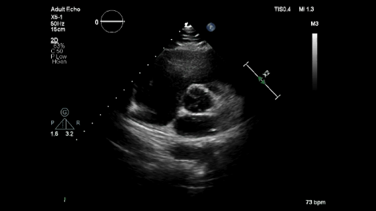
Parasternal short axis (PSAX)

The right ventricle appears enlarged in the parasternal short axis view. During acute right ventricular pressure load, the high pressure in the rightricle compresses the interventricular septum, causing a characteristic LV-D shape where both the left and right ventricles share the septum.
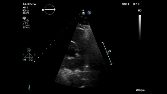
Apical four chamber (A4C)

The Apical four-chamber view image shows that the right ventricle is larger than the left ventricle in comparison.


CTPA

A thrombus in the shape of a saddle is lodged at the bifurcation of the pulmonary artery, as seen in the CTPA.

In the CTPA, the right ventricle enlarged in comparison to the left ventricle.
Management of RV failure in acute high risk acute pulmonary embolism

The left ventricle is perfused mainly during diastole. Because the LV produces a higher internal pressure during systole, systolic coronary resistance increases, and systolic coronary blood flow is lower.
The right ventricle receives equal perfusion during systole and diastole. However, in cases of acute PE, there is an acute increase in right ventricular systolic pressure (RVSP), which leads to right ventricular ischemia. Administering vasopressors can help increase blood pressure, restore coronary perfusion pressure gradient, and improve right ventricle inotropy.

Norepinephrine is commonly used to treat persistent hypotension by constricting blood vessels through α1—and β1 —adrenoceptors. Doses less than15 μg/min, NE has little to no effect on PVR. However, at higher doses, it may increase pulmonary vascular resistance (PVR).
Vasopressin, on the other hand, acts as a systemic vasoconstrictor and pulmonary vasodilator, making it a potential alternative to norepinephrine. Nonetheless, there is limited data on the use of vasopressin in patients with RHF.
Int J Cardiol. 2018 Dec 1;272S:46-52.
Risk stratification
PESI score (Pulmonary Embolism Severity Index)
Class I: ≤65 points, very low 30-day mortality risk (01.6%)
Class II: 66 - 85 points, low mortality risk (1.73.5%)
Class III: 86 - 105 points, moderate mortality risk (3.2-7.1%) Class IV: 106 - 125 points, high mortality risk (4.0 - 11.4%) Class V: >125 points, very high mortality risk (10.0 - 24.5%)
0 points: 30-day mortality risk 1.0% (95% CI 0.0 - 2.1%)
1 ≥ point(s): 30-day mortality risk 10.9% (95% CI 8.5 - 13.2%)
For patients with PESI class III-IV or sPESI ≥ 1, the mortality risk is intermediate.
If both RV dysfunction on TTE or CTPA AND elevated troponin levels are present, the risk is intermediate-high.
If one of the above or none, the risk is intermediate-low.
Patients who have acute PE and are hemodynamically unstable are at a high risk.
For PESI class I-II and sPESI = 0
The mortality risk is low if there is no RV dysfunction on TTE or CTPA.
However, if there is RV dysfunction, a troponin test should be performed.
If the troponin test is positive, the risk is intermediate-high,
if the troponin test is negative, the risk is intermediate-low.
Treatment PE with hemodynamic instability
Thrombolytic
rtPA
-
rtPA 100 mg over 2 h
-
0.6 mg/kg over 15 min (maximum dose 50 mg)
Although not officially approved, the accelerated rtPA regimen for pulmonary embolism is occasionally utilized in cases of severe hemodynamic instability, such as cardiac arrest.
Streptokinase
-
Accelerated regimen: 1.5 million IU over 2 h
-
250 000 IU as a loading dose over 30 min, followed by 100 000 IU/h over 12-24 h
Urokinase
-
Accelerated regimen: 3 million IU over 2 h
-
4400 IU/kg as a loading dose over 10 min, followed by 4400 IU/kg/h over 12 - 24 h
Contraindication
Absolute
-
History of haemorrhagic stroke or stroke of unknown origin
-
Ischaemic stroke in previous 6 months
-
Central nervous system neoplasm
-
Major trauma, surgery, or head injury in previous 3 weeks
-
Bleeding diathesis
-
Active bleeding
Relative
-
Transient ischaemic attack in previous 6 months
-
Oral anticoagulation
-
Pregnancy or first post-partum week
-
Non-compressible puncture sites
-
Traumatic resuscitation
-
Refractory hypertension (systolic BP >180 mmHg)
-
Advanced liver disease
-
Infective endocarditis
-
Active peptic ulcer
Surgical embolectomy
Catheter direct treatment
Anticoagulant
Heparin IV
Bolus dose 80 U/kg (Maximum 10,000 U) then 18 U/kg (Maximum 1800U/kg/h)
bridge with oral anticoagulant.
Treatment PE without hemodynamic instability
Low molecular weight heparin
Enoxaparin
For CrCl ≥ 30 ml/min: Enoxaparin 1 mg/kg sc q 12 h or 1.5 mg/kg subcutaneously once daily
Prophylaxis: 40 mg sc once daily
For CrCl < 30 ml/min: Enoxaparin 1 mg/kg sc once daily
Prophylaxis: 30 mg sc once daily
Fondaparinux
For CrCl ≥ 30 ml/min: Fondaparinux 5 mg sc daily (Wt < 50 kg)
Fondaparinux 7.5 mg sc daily (Wt 50-100 kg)
Fondaparinux 10 mg sc daily (Wt >100 kg)
Prophylaxis: 2.5 mg sc daily
For CrCl < 30 ml/min: Contraindicated

References
1) Konstantinides SV, Meyer G, Becattini C, Bueno H, Geersing GJ, Harjola VP, et al. 2019 ESC Guidelines for the diagnosis and management of acute pulmonary embolism developed in collaboration with the European Respiratory Society (ERS): The Task Force for the diagnosis and management of acute pulmonary embolism of the European Society of Cardiology (ESC). European Heart Journal. 2020 Jan 21;41(4):543–603.
2) Ramos H R, Ceballos M, Alvarenga H, et al. (September 26, 2019) Catheter-based Therapy for Massive Pulmonary Embolism in an Elderly Woman with Chest Pain and Dyspnea: Case Report. Cureus 11(9): e5771. doi:10.7759/cureus.5771
3) Sathe P, Patwa U. D Dimer in acute care. International journal of critical illness and injury science. 2014 Jul 1;4:229–32.
4) Olsson KM, Halank M, Egenlauf B, Fistera D, Gall H, Kaehler C, Kortmann K, Kramm T, Lichtblau M, Marra AM, Nagel C, Sablotzki A, Seyfarth HJ, Schranz D, Ulrich S, Hoeper MM, Lange TJ. Decompensated right heart failure, intensive care and perioperative management in patients with pulmonary hypertension: Updated recommendations from the Cologne Consensus Conference 2018. Int J Cardiol. 2018 Dec 1;272S:46-52. doi: 10.1016/j.ijcard.2018.08.081. Epub 2018 Aug 26. PMID: 30190155.
5) European Medicines Agency. (n.d.). *Arixtra: EPAR-product information* [PDF]. EMA. https://www.ema.europa.eu/en/documents/product-information/arixtra-epar-product-information_en.pdf
6) Patel R. Effective management of venous thromboembolism in the community: non-vitamin K antagonist oral anticoagulants. International Journal of General Medicine. 2016 May 4;9:107. doi:10.2147/IJGM.S100299