Mitral valve regurgitation
Echocardiography
Mitral valve prolapse
Parasternal long axis at end-systole shows a displacement of the posterior leaflets greater than 2 mm (indicated by the pink arrow) above the annulus plane.

AML
PML
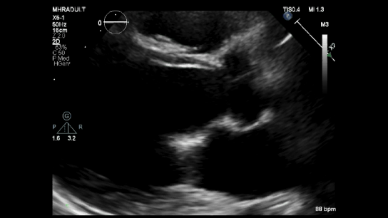
An M-mode echocardiogram from the parasternal long axis demonstrates the "Hammock sign," characterized by a U-shaped abnormal systolic motion and two distinct echoes during systole, representing the anterior and posterior leaflets.
Effective regurgitation orifice & MR volume measurement
Flow1 = Area x velocity = 4/2 π r² x Alias velocity
Flow2 = Area x velocity = ERO x MV Vmax
Flow1 = Flow2
4/2 π r² x Alias velocity = ERO x Peak MR velocity
2 π r² x Alias velocity = ERO x Peak MR velocity
ERO = 2 π r² x Alias velocity / Peak MR velocity
MR volume = ERO x MR VTI


References
O’Rourke R, Crawford M, Johnson A, Davidson R, LeWinter M, Karliner J. Prolapsing mitral valve leaflet syndrome. The Western journal of medicine. 1975 Apr 1;122:217–31.
Zoghbi, W. A., MD, FASE, & Adams, D., RCS, RDCS, FASE. (2017). Recommendations for Noninvasive Evaluation of Native Valvular Regurgitation. JASE, 30, 4th ser., 1-69. Retrieved June 12, 2017. DOI: 10.1016/j.echo.2017.01.007